

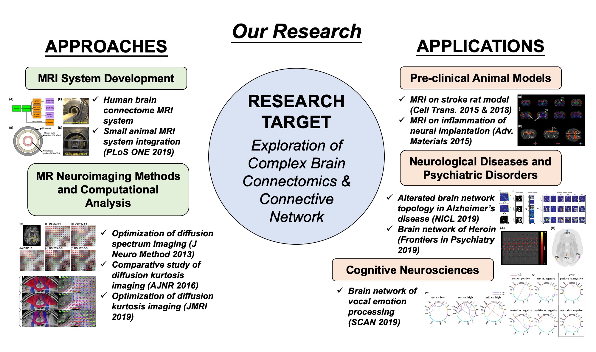
MRI System Development
The first aim of our research is to develop a multi-scale and high-performance MRI system for improving the angular and spatiotemporal resolutions to reveal finer details of the brain connectomics. This advanced MRI system incorporates two high-strength gradient coils with two different inner diameters, i.e. 48 and 12 cm, for imaging human and small animal, corresponding to gradient strengths of 150 and 675 mT/m, respectively. During the past few years, we have successfully established the multi-scale high-performance 3 T MRI system at NHRI.
Selected publications:
- Kuan-Hung Cho, Sheng-Min Huang, Chang-Hoon Choi, Ming-Jye Chen, Hsuan-Han Chiang, Richard P. Buschbeck, Ezequiel Farrher, N. Jon Shah, Ruslan Garipov, Ching-Ping Chang, Hsu Chang, Li-Wei Kuo. Development, integration and use of an ultra-high-strength gradient system on a human-size 3 T magnet for small animal MRI. PLoS One, 14(6):e0217916, June 2019, doi:10.1371/journal.pone.0217916.
MRI Neuroimaging Methods
The second aim of our research is to focus on developing MR neuroimaging methods and computational analysis. Specifically, we have focused on developing the novel high-angular resolution diffusion MRI methods and performed the optimization to improve the capability of resolving the complex tissue microstructures. We have optimized the diffusion kurtosis imaging, a novel technique to resolve tissue complex microstructures. Essentially, all these works would facilitate the applications on pre-clinical animal models and brain diseases in clinical settings.
Selected publications:
- Ezequiel Farrher, Farida Grinberg, Li-Wei Kuo, Kuan-Hung Cho, Richard Buschbeck, Ming-Jye Chen, Hsuan-Han Chiang, Chang-Hoon Choi, N. Jon Shah. Dedicated diffusion phantoms for the investigation of free water elimination and mapping: insights into the influence of T2 relaxation properties. NMR in Biomedicine, 33:e4210, April 2020, doi:10.1002/nbm.4210.
- Chia-Wen Chiang, Shih-Yen Lin, Kuan-Hung Cho, Kou-Jen Wu, Yun Wang and Li-Wei Kuo. “Effects of signal averaging, gradient encoding scheme, and spatial resolution on diffusion kurtosis imaging: An empirical study using 7T MRI,” Journal of Magnetic Resonance Imaging, 16 Apr 2019, doi: 10.1002/jmri.26755.
Computational Analysis Approaches
Graph-theoretical analysis has been developed as an important tool for investigating brain connective networks. Using graph-theoretical analysis to analyze the topological organization of brain structures and functions has been recently developed and considered as a new approach to explore the brain neuroscience. During the past few years, we have focused on optimizing the workflow of graph-theoretical analysis and further facilitating the use of graph-theoretical analysis on clinical applications. In our recent work, we investigated the alterations in brain network topology in the patients with mild cognitive impairment and Alzheimer’s disease. We also investigated the alteration of functional connectivity of default mode network in heroin users with different treatment modalities. Other than clinical applications, we also investigated the vocal emotion processing using the graph theoretical analysis.
Selected publications:
- Min-Chien Tu, Sheng-Min Huang, Yen-Hsuan Hsu, Jir-Jei Yang, Chien-Yuan Lin, Li-Wei Kuo. Discriminating Subcortical Ischemic Vascular Disease and Alzheimer’s Disease by Diffusion Kurtosis Imaging in Segregated Thalamic Regions. Human Brain Mapping, 1-14, January 2021, doi:10.1002/hbm.25342.
- Min-Chien Tu, Yen-Hsuan Hsu, Jir-Jei Yang, Wen-Hui Huang, Jie-Fu Deng, Shih-Yen Lin, Chien-Yuan Lin, Li-Wei Kuo. Attention and functional connectivity among patients with early-stage subcortical ischemic vascular disease and Alzheimer’s disease. Frontiers in Aging Neuroscience, 12:239, August 2020, doi:10.3389/fnagi.2020.00239.
- Li-Wei Kuo, Pei-Sheng Lin, Shih-Yen Lin, Ming-Fang Liu, Hengtai Jan, Hsin-Chien Lee, Sheng-Chang Wang. Functional Correlates of Resting-state Connectivity in the Default Mode Network of Heroin Users on Methadone Treatment and Medication-free Therapeutic Community Program. Frontiers in Psychiatry, 10:381, June 2019, doi:10.3389/fpsyt.2019.00381.
- Shih-Yen Lin, Chi-Chun Lee, Yong-Sheng Chen, Li-Wei Kuo. Investigation of functional brain network reconfiguration during vocal emotional processing using graph-theoretical analysis. Social Cognitive and Affective Neuroscience, 14(5):529-538, May 2019, doi:10.1093/scan/nsz025.
- Shih-Yen Lin, Chen-Pei Lin, Tsung-Jen Hsieh, Chung-Fen Lin, Sih-Huei Chen, Yi-Ping Chao, Yong-Sheng Chen, Chih-Cheng Hsu, Li-Wei Kuo. Multiparametric Graph Theoretical Analysis Reveals Altered Structural and Functional Network Topology in Alzheimer’s Disease. Neuroimage: Clinical, 22:101680, January 2019, doi:10.1016/j.nicl.2019.101680.